
Advancements in medical technology have made it possible to see inside the body with painless, noninvasive measures. These include x-rays and ultrasounds along with MRIs and CT scans. Each of these works in different ways and has different applications.
However, one thing remains consistent across each of these types of diagnostic imaging: they are safer, easier, and more comfortable than invasive measures. Therefore, even though it can be daunting when your doctor says you need one of these types of imaging, rest assured they will often be far simpler than patients expect.
Here we will look at the differences between x-rays and ultrasounds and the best uses for each.
Main Difference Between X-Ray and Ultrasound
Though there are many differences between how an ultrasound and x-ray works and how they are performed, the main difference a patient should understand is what each is used for. Ultrasound is used primarily for diagnostic imaging of soft tissue while x-rays are primarily used to see bone and some dense tissue areas.
For example, if you have a broken bone, an x-ray will be used. However, if you have a sprain or injury to the muscle, an ultrasound will be ordered. For injuries impacting both bones and soft tissue, your doctor may order both.
What is an X-Ray and How Does it Work?
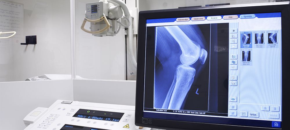
An x-ray is a type of diagnostic imaging that uses electromagnetic waves to get a picture of what is on the inside of your body. Specifically, it uses radiation to see through the layers of skin and other tissue in order to produce an image of your bones. This is possible because each part of your body absorbs x-ray energy at different rates.
So, while an x-ray may be able to pass through your skin, your bones are too dense. Therefore, the bones will show up white on an x-ray image while lungs, heart, and other organs will be dark. With this method, your doctor will be able to see breaks, fractures, and other abnormalities in your bones. Similarly, foreign objects will often be visible on an x-ray, making it an important tool for locating and removing metal and other materials that may become lodged in the body.
Are X-Rays Safe?
X-rays do expose you to radiation, however; the amount is typically quite small. For example, a chest x-ray exposes a patient to around the same amount of radiation you would naturally receive from the environment over a week and a half. Though the idea of radiation can be alarming, it is important to remember that radiation is already part of our environment. Moreover, the findings available from an x-ray can save you from surgery, make a surgery more effective, and help to catch health problems early.
What are X-Rays Used For?
X-rays, as mentioned above, are most often used to view bone fractures and breaks. However, they are also used for imaging for the following:
- Detecting certain cancers
- Locating bone tumors
- Dental imaging
- Swallowing difficulty (dysphagia)
- Lung problems
- Certain heart conditions
- Imaging to assist surgeons in certain procedures
What is an Ultrasound and How Does it Work?
An ultrasound, also called sonography, uses soundwaves to produce an image of your internal organs and other soft tissues. These sound waves are produced by a device called a transducer. This device sends out soundwaves and can also sense the sounds echoed back. Through these echoes, an image is formed of the area being observed.
Most often, a transducer is placed on the skin in order to produce an image of the tissues underneath. However, for certain conditions and issues and to produce the highest quality images, probes may be put on the inside of the body as well.

What is the Gel in Ultrasounds For?
If you have ever had an ultrasound or seen one performed, you have likely seen the technician spread a kind of gel on the skin before beginning the procedure. This gel creates a better seal between your skin and the ultrasound device, preventing any air pockets. This is necessary since ultrasound waves may be impeded by air.
Are Ultrasounds Safe?’
3D ultrasound imaging was first invented in the 1980s and became increasingly sophisticated. By the 90s it had become fairly common and was frequently used to assess pregnancy health. In the decades since its inception, there have been no risks detected.
Why Would You Need an Ultrasound?
Ultrasounds are used for a wide range of reasons, including both diagnostic reasons and therapeutic. It is most commonly used to check the health of a developing fetus since it does not pose a radiation concern like an x-ray does. These are some other common uses for ultrasounds:
- Detect thyroid issues
- Examine tumors
- Assess inflammation of the joints
- Assist in a prostate exam
- Check blood flow through the veins
- Detect gallbladder disease
Note, while ultrasounds are useful they do have significant limitations. Along with not being a good tool for producing images of bones, it also cannot take images of areas filled with air, such as the lungs. Just as a gel is needed to prevent air from forming between the skin and the transducer, the air in the lungs will block soundwaves.
CT Scans and MRIs
CT scans and MRIs are the other common diagnostic imaging tools. With an MRI (Magnetic Resonance Imaging) a magnetic field along with radio frequencies are used to produce a full body image. Your doctor may order this for a number of reasons as it is one of the best ways to get a total body scan of your internal organs and skeletal structure.
MRIs are commonly used to diagnose and assess issues with joints, the brain, heart, and blood vessels.
CT scans (Computed Tomography) takes a series of x-ray images to produce an image of everything from soft tissues to skeletal structure and even blood vessels.
CT scans are commonly used to diagnose and assess issues with bone fractures, tumors, and internal bleeding.
Both CT Scans and MRIs are less common than ultrasounds and x-rays. Both produce a more complete image of internal structures, but this may not be necessary in all situations. This is why your doctor will not order one unless necessary.
Where Can I Get Diagnostic Imaging?
Diagnostic imaging is an important part of diagnosing and treating various conditions. At BCML we offer each of these imaging services for your health and convenience. To schedule your appointment, call our caring team at 416-929-1900.