Seasonal sniffles, unexplained rashes, and late-night coughing fits share one root cause more often than you may think: allergies. An accurate test unpacks those symptoms, matches them to specific triggers, and lays the groundwork for a care plan you can trust.
At downtown Toronto’s Lockwood Clinic, allergy testing fits smoothly into a single visit, giving you fast answers and a clear path forward.
Related Article: Services You Can Expect at a Walk-In Clinic in Toronto
What Is an Allergy?
An allergy is an exaggerated immune response to a harmless substance such as pollen, dust mites, or certain foods. When your body mistakes that substance for a threat, it releases histamine and other chemicals that create itch, swelling, sneezing, or wheezing. Pinpointing the exact trigger allows you to limit exposure and choose treatments that calm the immune system instead of masking flare-ups.
Why Consider Allergy Testing?
Most people reach for over-the-counter pills when symptoms strike, yet guessing the trigger can keep you in a cycle of flare and relief. Testing provides:
- Clarity: Know whether the culprit is ragweed, cat dander, or last night’s shrimp.
- Targeted care: Select antihistamines, nasal sprays, or immunotherapy that match the trigger.
- Lifestyle tips: Adjust cleaning routines, air filters, or meal plans based on solid evidence.
- Peace of mind: Rule out serious conditions that can mimic allergy signs, such as asthma or eczema.
Types of Allergy Tests Offered at Lockwood Clinic
Before any samples are taken, your clinician will help you match the test to your symptoms, medical history, and medication list. Having the right method from the start avoids repeat appointments and gives the most reliable answers.
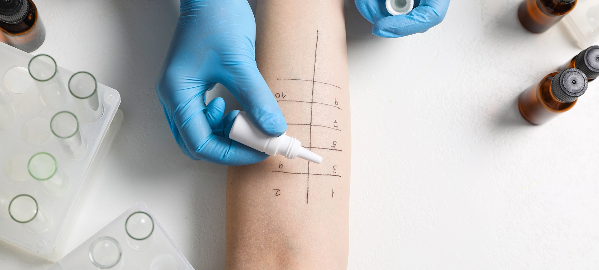
- Skin Prick Test: A nurse places tiny drops of diluted allergens (e.g. as ragweed, mould, or pet dander) on your forearm or upper back, then makes a shallow scratch through each drop. If you are sensitive, a small, mosquito-bite-sized welt appears within 15 minutes. This test covers up to 40 allergens at once and is usually painless, aside from mild itching.
- Intradermal Test: When a skin-prick result is borderline or when drug allergies are suspected, a trace amount of the allergen is injected just under the first skin layer. Because it reaches slightly deeper tissue, this method picks up reactions the prick test can miss.
- Patch Test: Ideal for contact dermatitis, the test uses adhesive patches containing common culprits (e.g. nickel, latex, fragrances) that stay on your back for 48 hours. After removal, the nurse checks for redness or blistering that signals sensitivity to products that touch your skin.
- Specific IgE Blood Test: A single blood draw measures the level of allergy antibodies circulating in your system. It is perfect for patients who cannot stop antihistamines, have eczema that makes skin tests hard to read, or want results for dozens of allergens without multiple needle pricks.

How to Prepare for Your Appointment
A smooth session starts with a little planning.
- Pause antihistamines: Stop oral and topical antihistamines five days before testing so they do not blunt the skin response.
- Gather a symptom diary: Note when flare-ups happen, what you ate, and where you were. The diary guides test selection.
- Wear short sleeves or loose clothing: Easy access speeds the skin test process.
- Eat a light meal: A steady blood sugar level makes blood draws easier and keeps you comfortable.
What Happens on Test Day?
Each step is quick, gentle, and explained in plain language.
- Check-in and medical history review: A nurse confirms medications, recent illnesses, and past reactions.
- Skin preparation: The test area is cleaned with alcohol to prevent infection.
- Allergen application: Drops or injections go on marked spots. You may feel mild pressure but no real pain.
- Observation period: You relax in the clinic lounge while staff monitor for bumps or redness.
- Measurement and recording: Raised areas are measured in millimetres to grade reaction strength.
- Blood draw (if needed): A phlebotomist collects a small sample for laboratory analysis.
- Results discussion: A clinician explains findings, answers questions, and outlines next steps. You leave with printed results and a care plan.
The entire visit usually runs 60 to 90 minutes, and you can resume normal activities right after.
Understanding Your Results
Numbers and millimetre readings only matter when they translate into day-to-day choices. Your clinician will pair the data with your symptom diary to build a practical action plan.
- Positive Result: A raised bump on skin testing or an elevated IgE level confirms that your immune system flags that substance as a threat. Expect advice on targeted medications, avoidance tactics, and possible immunotherapy.
- Negative Result: No measurable reaction means that the allergen is unlikely to be driving your symptoms. Your clinician may investigate other triggers, such as irritants or chronic sinus issues.
- Borderline Result: A mild skin welt or mid-range antibody count can go either way. Follow-up options include a repeat test in several months, an elimination diet, or a monitored exposure challenge to settle the question.
How Allergy Testing Improves Daily Living
Knowing the trigger is only the first step; acting on it brings the real payoff.
- Medication match: Choose the right antihistamine or inhaler strength and avoid drug side effects from guesswork.
- Home adjustments: Install high-efficiency filters, wash bedding in hot water weekly, or keep pets out of bedrooms.
- Diet upgrades: Swap allergenic foods for safe, nutritious options and remove the fear of accidental reactions.
- Immunotherapy pathway: Candidates with severe or multiple allergies can start shots or tablets that retrain the immune system over time.
- Travel confidence: Pack wisely for trips, avoiding unknown foods or hotel rooms with down bedding.

Common Myths About Allergy Testing
Misconceptions stop many people from seeking the relief they deserve. Let’s clear up the biggest ones.
- “Testing is painful.” Skin pricks feel like light scratches, and blood draws use a fine-gauge needle. Most patients describe mild, momentary discomfort.
- “It takes forever to get answers.” Skin-test reactions appear in 15 minutes, and blood-work results usually come back within a week.
- “Children are too young to be tested.” Infants as young as six months can be assessed safely, allowing parents to adjust diets or home environments early on.
- “Drugstore tablets are all I need.” Antihistamines mask symptoms but do not tell you what to avoid or whether immunotherapy could cut your medication use long-term.
Related Article: Understanding the Benefits of Family Practice: Comprehensive Healthcare for All Ages
Why Choose Lockwood Clinic for Allergy Testing?
Identifying a trigger is only half the battle; you also want a walk-in clinic that makes the whole experience seamless. Lockwood Clinic combines nearly a century of medical expertise with modern walk-in convenience, letting you move from testing to treatment without extra stops.
- One-Stop Care: Family physicians, diagnostic nurses, and an on-site pharmacy share one roof, so you can consult, test, and fill prescriptions in a single visit.
- Same-Day Access: Walk-in and extended-hour appointments mean you avoid weeks-long waits common at many specialty centres.
- Downtown Location: Situated close to multiple transit lines and with parking nearby, the clinic is easy to reach whether you live in the core or commute in for work.
- Experienced Team: Decades of collective experience ensure precise testing techniques and clear explanations. No medical jargon, just actionable advice.
- Flexible Follow-Up: Prefer virtual check-ins? Need an after-work slot? The clinic offers phone, video, and late-day appointments to keep your care on track without disrupting your schedule.
Related Article: Benefits of Toronto’s Walk-In Clinics: Fast, Flexible, and OHIP-Covered Care
Book Your Allergy Assessment
Clear breathing, restful sleep, and rash-free skin start with solid information. Schedule an allergy test at Lockwood Clinic, bring your symptom diary, and leave with a concrete plan to feel better. Call, walk in, or book online today; relief is closer than you think.